This blog on the biology of insulin is part of a series celebrating the 1ooth anniversary of the discovery of insulin. You can check out previous blogs in this series, written by CIM Intern Eleanor Medley, here!
If you have diabetes, you probably know what insulin looks like, feels like, even smells like,1 but what does it do, exactly? Here’s a quick rundown of the structure and function of insulin.
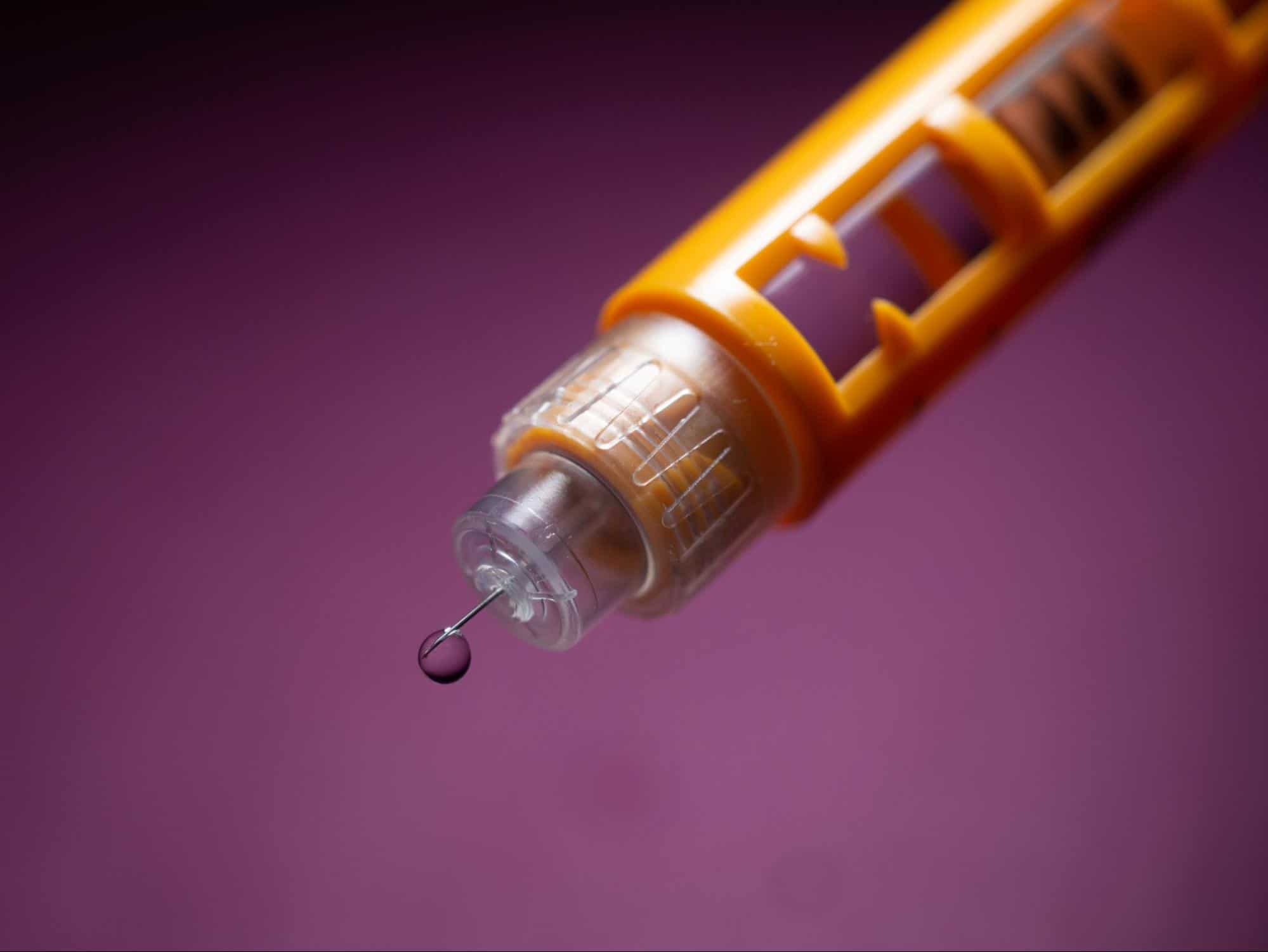
Novolog insulin pen, 4mm bd nano needle with a drop of insulin.
At a Glance
Insulin is a hormone produced by beta cells in the pancreas. It is secreted when BG levels rise and allows the body to utilize glucose for energy.
Gene
Humans have a single insulin gene, called INS. It is located on Chromosome 11.2
Synthesis
Insulin is made by beta cells in a part of the pancreas called the islets of Langerhans. First, the insulin gene is transcribed and translated into a precursor called preproinsulin which then gets converted to proinsulin. One of the protein chains, the ‘C-peptide’, then gets excised, creating the mature form of insulin.3
Protein Structure
Mature insulin is a protein composed of two chains, A and B. They are linked together through bridges connecting sulfur atoms.4 Individual insulin molecules tend to come together in groups of 2, or 6 when zinc is present.5
Secretion
Beta cells store and secrete insulin in response to particular signals. The most important signal for insulin secretion is increasing BG levels in the blood (following a meal, for example).6 Insulin secretion can also be stimulated by certain amino acids, fatty acids, and keto acids.
The secretion of insulin can be inhibited when the sympathetic nervous system responsible for the fight-or-flight response is activated.7
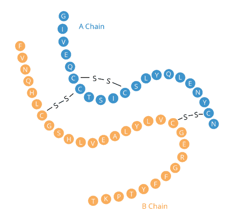
Blue circles coded with letters representing amino acids making up insulin’s A chain. Orange circles coded with letters representing amino acids making up insulin’s B chain. A disulfide bridge connects two amino acids in the A chain. Two more disulfide bridges connect the A chain to the B chain.
Function
Once secreted, insulin binds to specific receptors on the outside of target cells. This moves glucose transporters to the surface of these cells, allowing them to take in glucose and use it for energy.8 In this way, insulin can be thought of as a key to unlock doors allowing glucose into cells.
Insulin stimulates glucose uptake by fat, muscle, and liver tissue.9 In muscles, glucose is stored as glycogen which can be broken down for energy during exercise or fasting.
References
References
[1] Wardian, Jana L. “The Smell of Diabetes.” Clinical Diabetes, vol. 36, no. 3, 2018, pp. 257–258., doi:10.2337/cd18-0010.
[2] Tokarz, Victoria L et al. “The cell biology of systemic insulin function.” The Journal of cell biology vol. 217,7 (2018): 2273-2289. doi:10.1083/jcb.201802095
[3] “Insulin – Structure – Function.” TeachMePhysiology, 8 July 2020, teachmephysiology.com/endocrine-system/pancreas/insulin/.
[4] Utiger, Robert D. “Insulin.” Encyclopædia Britannica, Encyclopædia Britannica, Inc., 13 Mar. 2020, www.britannica.com/science/insulin.
[5] “Structure of Insulin.” Vivo Pathophysiology, www.vivo.colostate.edu/hbooks/pathphys/endocrine/pancreas/insulin_struct.html.
[6] Utiger, “Insulin”
[7] Utiger, “Insulin”
[8] Utiger, “Insulin”
[9] Utiger, “Insulin”